
Using CT scanners to investigate bees. Dr Mark Greco
Using CT scanners to investigate bees. Dr Mark Greco
For a period of 2 years, between 2015 and 2017, the Eva Crane Trust supported some of the research being carried out by Dr Mark Greco at Charles Strut University, Australia.
Dr Greco investigated the presence and spread of pathogens in honeybee colonies which has traditionally been assessed using crude invasive manual methods such as opening the hive and visually inspecting colonies for signs of disease. Research has shown that many pathogens affect honeybee health; however, Dr Greco explains that, ‘there is still much that we do not know on how these pathogens are spread from one honeybee to another, one hive to another or one apiary to another’.
Dr Greco believes that: ‘This research will develop a more powerful, accurate and non-invasive method for diagnosing pathogen dispersal within honeybee colonies. A tracking system, using X-ray Computerised Tomography (CT), will be developed to show how and when honeybees pass food from one bee to another within the hive. Human body CT scanners will be used to visualise the inside of honeybee hives. The food will be labelled and will contain p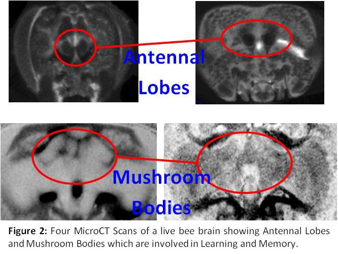 athogen so that the 3-D X-ray imaging produced by the CT scanning can track how and when the pathogen is spread within the hive. The method will also be valuable for other insect pollinators such as bumble bees, solitary bees, ants, wasps and also for the commercially important species of termites’.
https://link.springer.com/article/10.1007/s00040-015-0432-4
No spatial patterns for early nectar storage in honey bee colonies
Eyer, M., Greco, M.K., Lang, J. et al. Insect. Soc. (2016) 63: 51. https://doi.org/10.1007/s00040-015-0432-4
Abstract
Honey bees, Apis, forage for nectar and pollen, which are subsequently stored in cells of their nests. Despite the importance of honey storage for colony survival, very little is known about decision making by honey bee workers that could optimise the transformation of nectar into honey. Here we test, using diagnostic radioentomology, whether workers use rules based on sugar concentration to optimise the spatial distribution of storage cells during nectar ripening. The data show that after the first 3 days of storing activity, various sugar concentrations were mixed in individual cells. A spatial clustering of cells with content of similar concentration was only occasionally observed. The results, therefore, suggest that at early stages of storage, spatial proximity of cells with similar sugar concentrations does not result in improved efficiency and, therefore, does not seem adaptive. The costs involved in locating particular cells probably outweighs the benefits of clustering. Alternatively, but not mutually exclusive, physiological constraints (e.g. variation in the perception of sugar concentration) might limit such optimisation behaviour. Storing behaviour can serve as a model to better understand food provisioning and complex organisation of insect societies.
Article: http://iopscience.iop.org/article/10.1088/2057-1976/aa6307/meta
Applying x-ray micro-tomography to learning and memory
Mark K Greco and Timothy Stait-Gardner. Biomedical Physics & Engineering Express, Vol. 3 (2) March 2017
Abstract
Metabolic processes and neural pathways such as those for glucose metabolism and olfaction in insects are similar to, and in some cases identical to, those found in vertebrates. In particular, the peripheral architecture of the insect and mammalian olfactory system is surprisingly similar. Thus one insect, the western honeybee (Apis mellifera), has become a model organism for the investigation of olfactory signal processing in the brain. A. Mellifera has superior abiliti
es to identify, classify, learn and remember odoriferous environmental chemicals. The use of x-ray MicroCT imaging for the non-invasive study of insects (termed diagnostic radioentomology 'DR') is increasing. This experiment was conducted as a 'proof of principle' study to identify whether DR scanning live insects (honeybees) can be used to non-invasively examine learning and memory with respect to brain architecture and function. A bench-top MicroCT scanner was used to scan live bees. To enhance tissue differentiation, radiographic contrast was injected directly into the haemolymph. Brain volume was measured using BeeView software. Gross brain architecture was visualised in 2D and 3D projections. Scanning of live bees enabled minimally-invasive imaging of physiological processes for the first time such as passage of contrast from gut to haemolymph as well as preliminary brain perfusion studies. In particular, the learning and memory neuropils in the antennal lobes were assessed by measuring changes in antennal lobe volumes. Future experiments will correlate neuropil volume changes with classic PER learning and memory studies. One advantage of using an insect model is that multiple experiments can be conducted without the need for lengthy ethics approvals that apply to vertebrate experiments. The advantage of using live insects is that longitudinal studies can be performed on individuals therefore reducing, to a great extent, the inherent inter-cohort errors which occur. Once proof of principles is established in the insect model, experiments such as these can easily be extended to vertebrates.
The funding has allowed him to analyse thousands of data files from previous experiments and use the information in his latest peer reviewed article:
Exposure of Insects to Radio- Frequency Electromagnetic Fields from 2 to 120 GHz
Arno Thielens1, Duncan Bell, David B. Mortimor, Mark K. Greco, Luc Martens & Wout Joseph
NATURE: Scientific Report, March 2018.
Abstract
Insects are continually exposed to Radio-Frequency (RF) electromagnetic fields at different frequencies. The range of frequencies used for wireless telecommunication systems will increase in the near future from below 6 GHz (2 G, 3 G, 4 G, and WiFi) to frequencies up to 120 GHz (5 G). This paper is the first to report the absorbed RF electromagnetic power in four different types of insects as a function of frequency from 2 GHz to 120 GHz. A set of insect models was obtained using novel Micro-CT (computer tomography) imaging. These models were used for the first time in finite-difference time-domain electromagnetic simulations. All insects showed a dependence of the absorbed power on the frequency.
All insects showed a general increase in absorbed RF power at and above 6 GHz, in comparison to the absorbed RF power below 6 GHz. Our simulations showed that a shift of 10% of the incident power density to frequencies above 6 GHz would lead to an increase in absorbed power between 3–370%.
More information about Dr Greco’s work can also be found on our News page.